Specifications:
|
Display model |
Separated |
|
Optical quality |
Standard |
|
Field of view |
Maximum 53° |
|
NO pupil scatter photographing |
support |
|
Minimum pupil diameter |
3.3mm |
|
Color digital collector |
≧12M pixel |
|
Black and white collector |
≧1300 line |
|
Radiography method |
Flash radiography |
|
|
Dynamic radiography videoing |
|
Observation illumination |
Infrared light |
|
Refraction compensation range |
≧±15D |
|
Outside fixed viewing target |
|
|
Single point fixed viewing target |
|
|
Auxiliary para-position |
Double point auxiliary focusing |
|
Focusing method |
Manual |
|
Exposure method |
Manual |
|
Medical internet |
Dicom 3.0 interface |
|
Second display screen |
Optional |
|
Eye position recognition |
Automatic |
|
Optical incline angle |
Horizontal ±30° Vertical ±12.5° |
|
Working distance |
42mm±2mm |
|
Operation stage |
Standard |
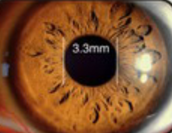
Minimum
pupil photograph diameter3.3mm
More
patients can be checked
Applicable
for ophthalmology section use,
physical
examination, diabetes patients fundus check.
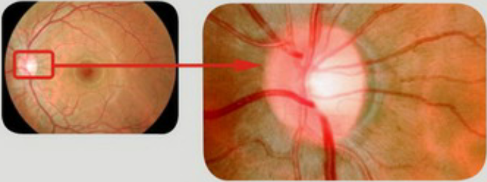
Inner
fixed digital collector Offer12Mpixel high definition color fundus
image
High resolution radiography image
Can clearly view Macular arch ring and
other Small vessels tiny nidus, good for laser treatment.
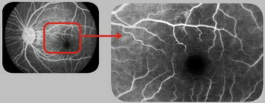
Field
of view single picture can reach maximum 53°
Assure
precise fundus checking, avoiding missed diagnosis ratio, elevating checking
ratio
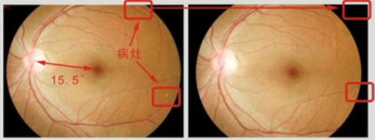
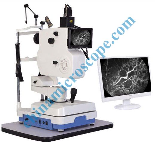
Double
radiography model
1. Flash radiography, infrared light
observation, flash illumination, not dazzling for patients with good
cooperation
2. Continuous dynamic radiography, can record
early full-fill process
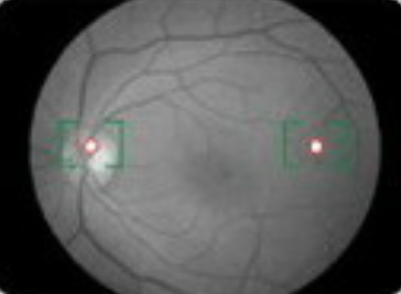
Double point auxiliary focusing
Easy Eye position recognition, you just
need to make the two light spots in the screen to the maximum.
Optical
incline with inner, exterior fixed viewing target, can photographing
180°fundus
Optical
incline angle Horizontal
±30° Vertical
±12.5°
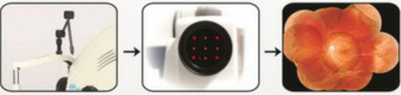
Newly
designed five points, nine points inner fixed viewing target, guiding the eye
movement, shooting from
all angle