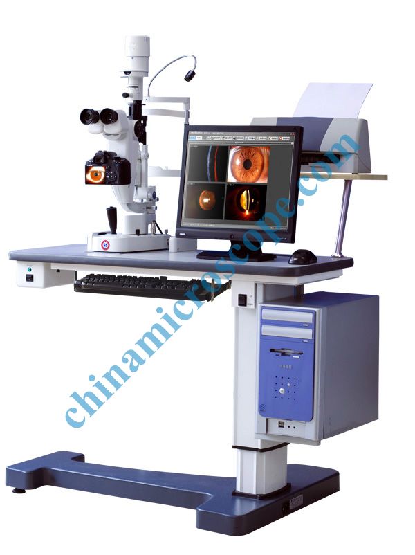
Characteristics:
•What doctors see from eyepieces is showed synchronously on camera display screen, which is very convenient for the doctors to capture the best and clearest pictures.
•Photos taken by the 12.4 megapixel camera.
•Background illumination specifically designed for darkroom environment.
•Fine photos immediately taken by the button on joystick.
•Used for establishing the full data of patients, processing images, measuring the length, area and so on.
Specifications:
|
Microscope Type |
Galilean stereoscopic microscope |
|
|
Magnification |
Five-step drum magnifications |
|
|
Eyepiece |
12.5X |
|
|
Magnification Ratio(Field of View) |
6 x (Æ33) 10x(Æ22.5) 16x (Æ14) 25´ (Æ8.8) 40´(Æ5.5) |
|
|
Range ofPupil Distance |
45 mm ~ 75 mm |
|
|
Adjustment of Diopter |
-5D ~ +3D |
|
|
Slit Width |
0mm~14mm adjustable(slit is round when the slit width is14mm) |
|
|
Slit Height |
1mm~14mm adjustable |
|
|
Aperture Diameter |
φ14mm、φ10mm、φ5mm、φ2mm、φ1mm、φ0.2mm |
|
|
Slit Angle |
0°~180° adjustable |
|
|
Slit Inclination |
5°、10°、15°、20° |
|
|
Filter |
heat absorption, grey, redfree, cobalt blue |
|
|
Illumination Bulb |
12v30w Halogen Bulb |
|
|
Maxial Illuminance |
≥280000 Lx |
|
|
Preceding Mirror |
-58.7D |
|
|
Collection Medium |
Single Lens Glistening Camera |
3CCD |
|
Special Specifications |
Static Pixel:12.4 megapixel |
Dynamic Image:795×596×3 |
|
Double display screens |
Single display screen |
|
|
Without the function of kinescope |
With the function of kinescope |
|
|
Illuminations Type of Filming |
Background Light |
|
|
Functions of images processing. |
Measuring length, width, angle and area; adding arrowhead and letters; processing images. |
|
|
Assistant Diagnosis |
With the diagnosis database. |
|